


KEY FEATURES
Live Cell Observation
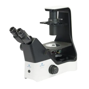
The EXI-310 is available with phase contrast observation for viewing live cells without the need for staining. The phase contrast configuration includes a phase contrast slider, centering telescope, a Plan 4X objective, and 10X and 20X Plan Phase objectives – 4X and 40X Plan Phase objectives are optional accessories. Add the optional mechanical stage and stage inserts to simplify sample movement and positioning.
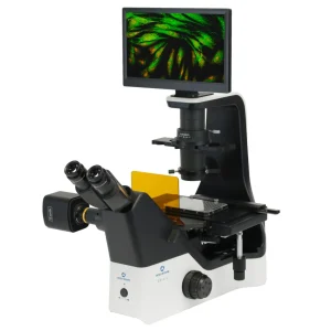
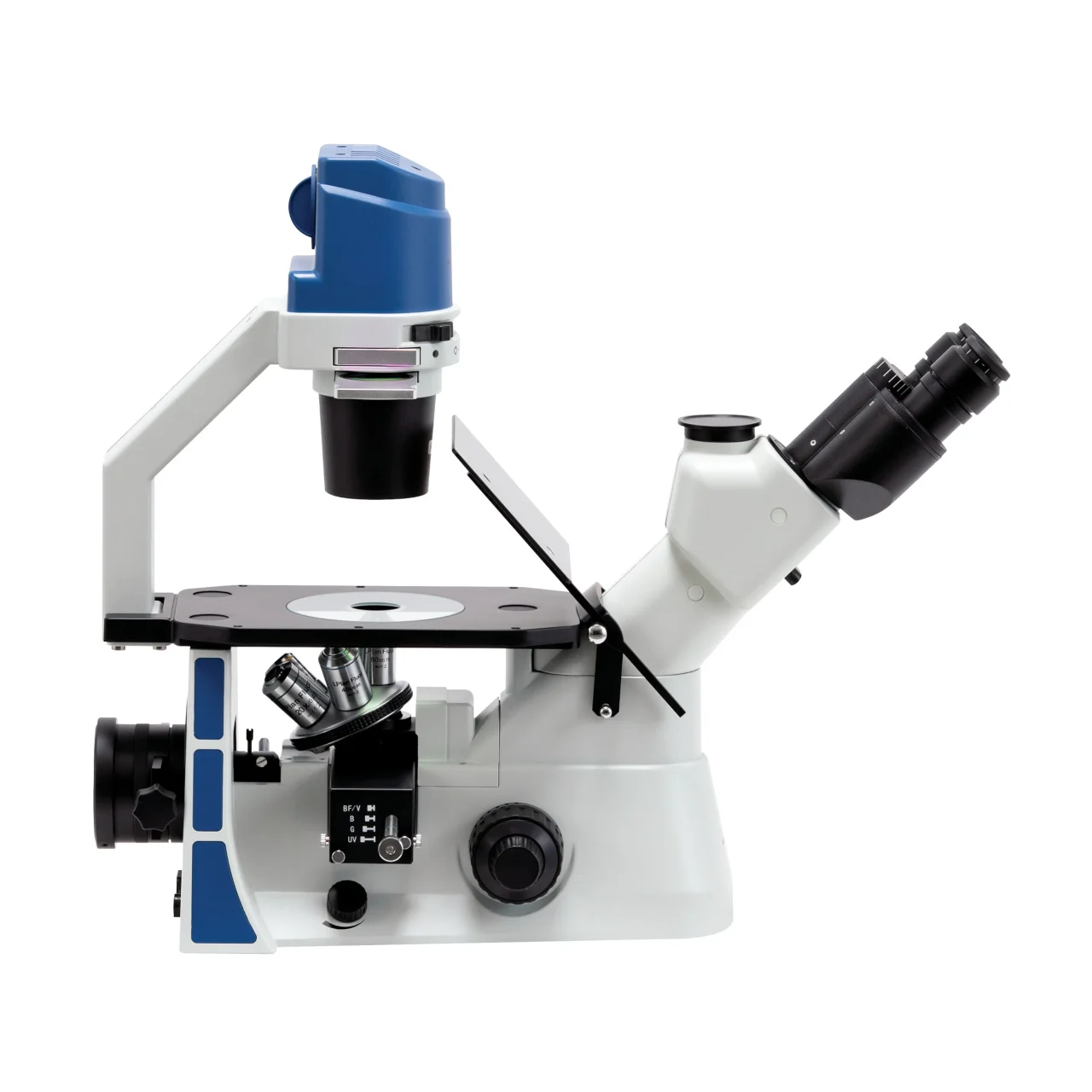
Fluorescence Observation
Available as a standard system solution or purchased as optional accessories, fluorescence observation expands the EXI-310 capabilities for observing live cells expressing fluorescent proteins or fixed cells stained to localize intracellular components. The epi-fluorescence illuminator of the EXI-310 accepts a variety of external solid state fluorescence light sources.
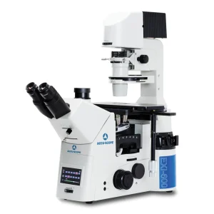
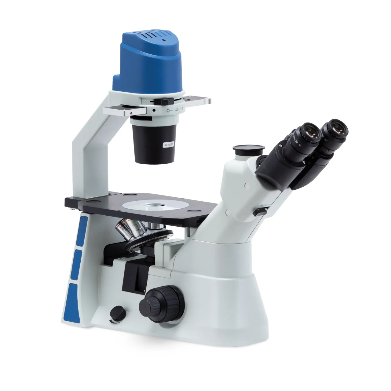
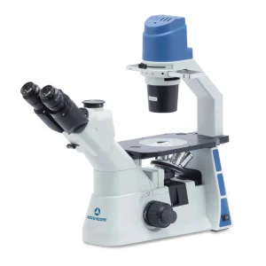
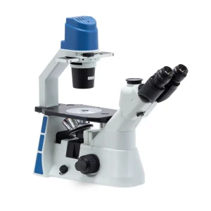
Professional Quality. Small Footprint.
Delivering outstanding image, the EXI-310 maintains a small footprint, leaving precious bench space for sample preparation or other lab instruments. Mount an optional camera to the top of the viewing head to permit unrestricted access to the focus knobs, illumination adjustment, or optional fluorescence slider or stage controls.
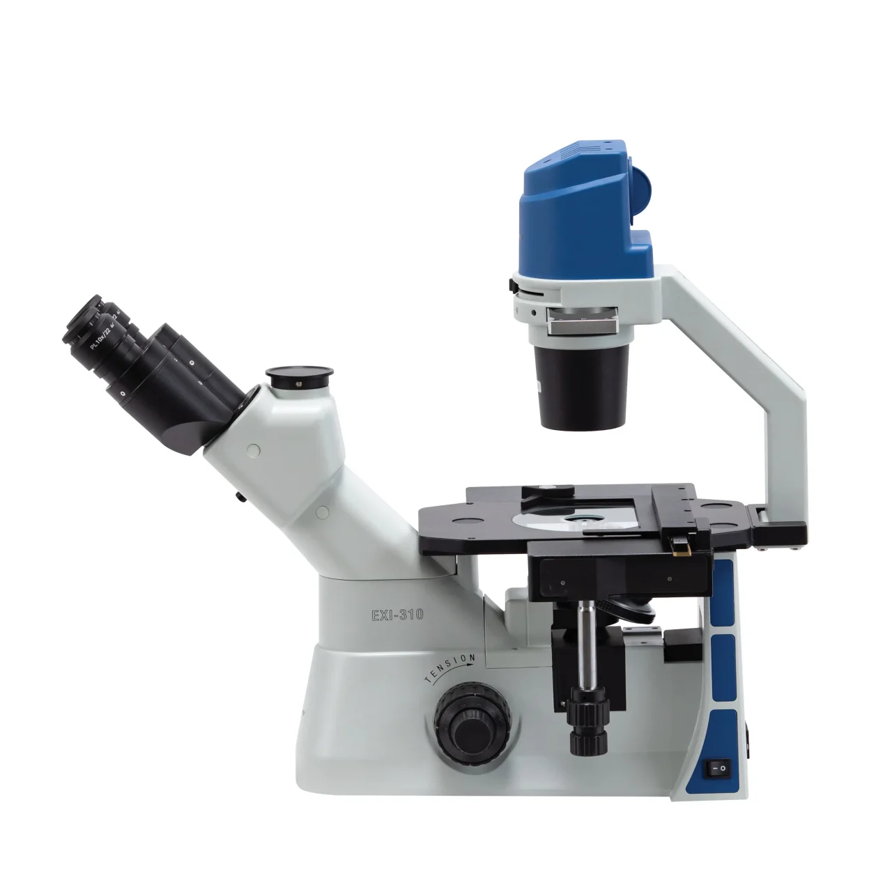
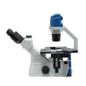
Specimen Positioning. Stage Configuration.
The standard flat stage on the EXI-310 allows fast sample loading and positioning during routine observation. Add the optional mechanical stage for more precise specimen positioning, often needed during observation at higher magnifications. The mechanical stage readily accepts multi-well plates, and a variety of stage inserts are available to accommodate Petri dishes, standard microscope slides and slide chambers, and Terasaki plates.
Optical Excellence
Defined by superb clarity, sharpness, and resolution, the EXI-310 objectives provide bright images for routine and challenging laboratory observation work. A wide range of objectives are available including Plan Phase, Plan Achromat, Plan Phase Fluor, and Plan Fluor.
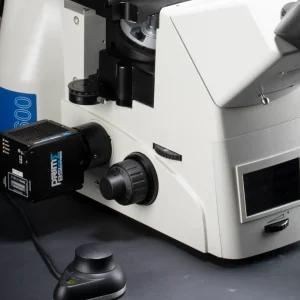
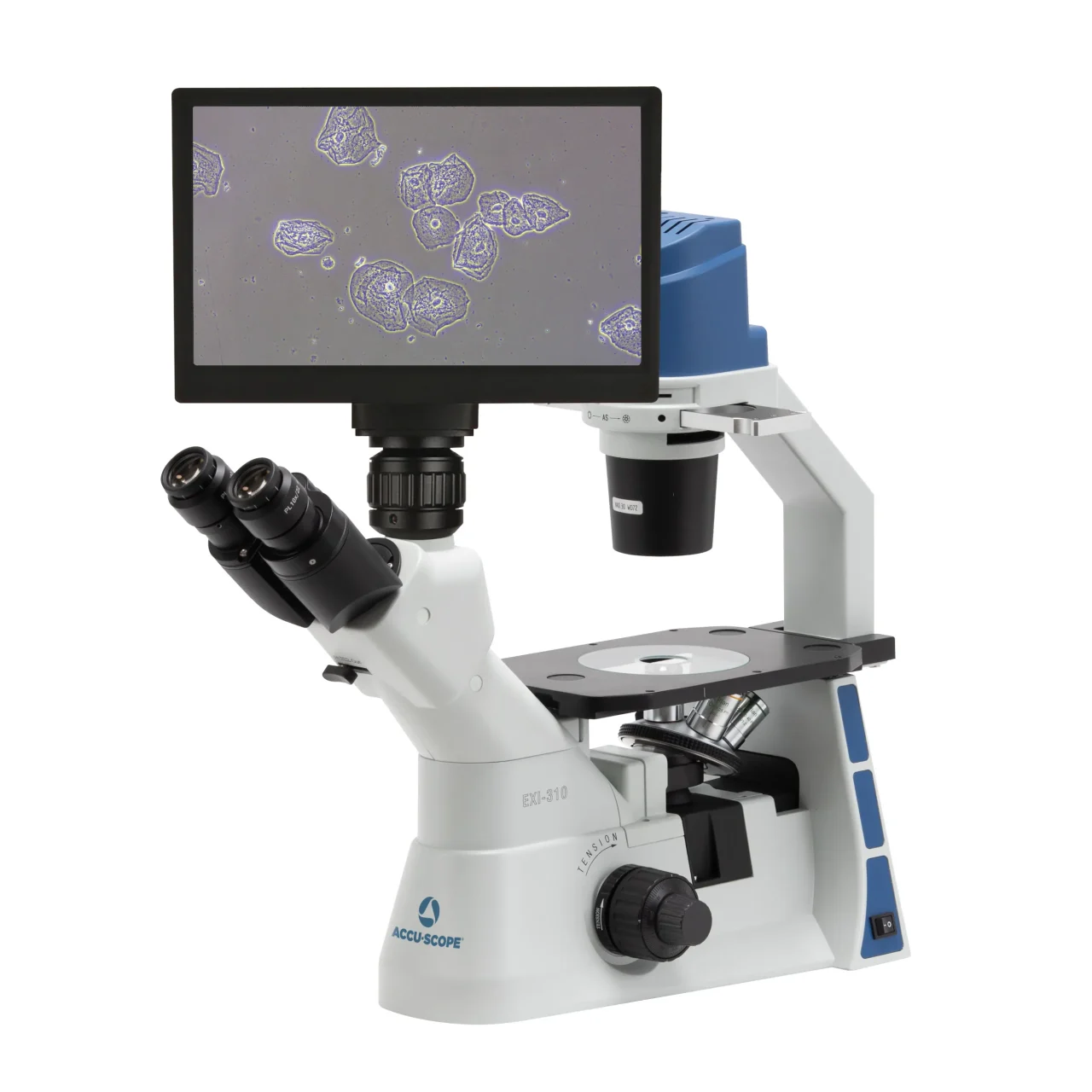
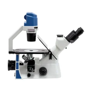
Digital Imaging
Digital images provide documentation, measurement and analysis of samples being studied. Monitors promote improved ergonomics during microscope use and allow for easy viewing of samples and collaboration with colleagues. Cameras mount to the top of the viewing head, permitting unrestricted access to the focus knobs, illumination control and the stage for sample positioning. Cameras and camera adapters are sold separately.